

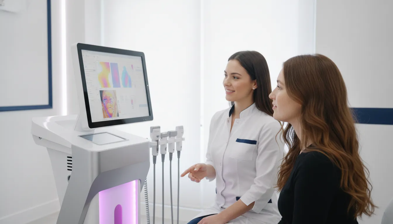
Professional practices often need more than a standard photograph to explain what a patient is seeing and what a consultation should consider. Consistent imaging can help teams document visible skin characteristics, compare images over time, and make conversations more specific without turning a picture into a diagnosis.
A skin scanner uses controlled imaging, and in some systems multiple light modes, to help visualize features such as texture, pores, wrinkles, pigmentation, vascularity, and hydration. Its output supports consultation, education, and documentation, while interpretation remains the responsibility of a qualified professional.
For practices evaluating this technology, the useful question is not simply how many images a system can produce. It is how consistently those images can be captured, understood, and incorporated into a patient-centered workflow. Exploring professional skin imaging systems starts with understanding what the equipment is designed to show and how those capabilities fit a practice.
What Is a Skin Scanner for Professional Practices?
A professional skin scanner is a controlled imaging system that captures standardized views of the face for visual assessment, consultation, and documentation. Unlike a single point measurement, full-face imaging is designed to show broader facial skin characteristics in a consistent format. A review in Frontiers in Medicine describes computer-assisted full facial skin imaging systems as widely used in dermatology and medical cosmetology. The systems can produce reproducible digital images and quantitative data for evaluation and follow-up. Read the review.
Depending on the system and model, a practice may capture images using standard, polarized, cross-polarized, or UV light. These different lighting conditions can help visualize surface features and selected below-surface patterns that may not be apparent in an ordinary photograph. Emage Medical’s ImagePro platform, for example, is positioned as an imaging and consultation technology for professional practices. Its listed analysis parameters include features such as pores, wrinkles, pigmentation, skin tone, vascularity or inflammation, texture, sebum levels, and hydration. The available parameters and lighting modes vary by model, so practices should evaluate the specific configuration rather than assume every skin scanner offers the same output.
How full-face imaging differs from consumer photo apps
A consumer photo app usually depends on a phone camera, uncontrolled room lighting, changing distance, and a user’s angle. Filters or algorithmic overlays may make an image look more detailed, but they do not necessarily create a repeatable record for a professional consultation. A dedicated skin scanner is built around a capture process that can standardize positioning, illumination, and views. That consistency gives a practice a stronger basis for comparing images over time and discussing what the patient can see.
Consistency still requires trained operation. Differences in environmental lighting or viewing angle can affect follow-up results. And the same academic review notes that familiarity with imaging principles is important for using the technique well. A skin scanner supports the conversation; it does not diagnose a condition or replace patient history, physical examination, or clinician judgment. Its role is to help practices explain visible findings, document the consultation, and make patient-specific discussions more concrete.
What Can a Professional Skin Scanner Reveal?
A professional skin scanner can help a practice visualize patterns that are difficult to compare through a quick visual inspection alone. Depending on the system and lighting modes, imaging may show surface characteristics such as pores, wrinkles. And texture, while specialized views can support assessment of pigmentation, vascularity, inflammation, hydration, and related parameters. These outputs are best used as visual and consultation tools, not as diagnoses.
For example, white-light imaging is commonly used to evaluate texture, wrinkles, and pores in studies of facial skin and aging-related treatments. A review of computer-assisted facial imaging also describes pigmentation, inflammation, vascular patterns, skin texture, and apparent aging severity as areas these systems may assess. Read the academic review of facial skin imaging systems for additional context.
Surface features and visible skin patterns
Standard views can provide a consistent way to discuss the appearance of pores, fine lines, wrinkles, uneven texture, and overall skin tone. This gives the clinician and patient a shared visual reference during a consultation. It can also support documentation over time, provided images are captured under comparable conditions. The lighting, camera angle, and operating technique matter because inconsistent follow-up conditions can change the apparent result.
Color, pigmentation, and vascular patterns
Specialized lighting can add another layer of visual information. Imaging systems described in the research use modes such as white light, UV, brown-spot, red-area, and UV-fluorescence views. Brown-spot views may present areas of hyperpigmentation, while red-area patterns may represent color concentrations associated with vascularity or inflammation. These visual patterns should be discussed carefully. They can support assessment and patient education, but they do not establish the cause of a finding or replace clinical judgment.
Emage Medical’s ImagePro overview lists pores, wrinkles, dermal and epidermal pigmentation, skin tone, skin type classification. Vascularity and inflammation, sebum levels, porphyrin ratio, texture, and hydration among its analysis parameters. The specific parameters and presentation depend on the model. Practices evaluating a professional skin imaging system should confirm which views and outputs are included in the configuration they are considering.
Hydration and related measurements can add useful context to a consultation, especially when paired with the patient’s history and the clinician’s examination. The most responsible interpretation combines the images with professional expertise, rather than treating a score, color map, or AI-assisted output as a standalone medical conclusion.
How Should Practices Interpret Skin Scanner Results?
A skin scanner is most useful when the practice treats its output as structured visual information, not as a standalone clinical conclusion. Staff can use images and measurements to organize a conversation, identify areas worth discussing, and document what was reviewed. The patient’s history, direct examination, goals, and the clinician’s judgment remain essential to interpretation.
Start with a consistent capture process
Before comparing results, make the capture conditions as consistent as possible. Use the same device settings, lighting approach, patient positioning, facial expression, and viewing angles whenever the practice repeats an image. Record relevant workflow details, such as whether the patient arrived with makeup or skincare products that could affect the visual presentation. This discipline makes changes easier to discuss without confusing a difference in technique with a difference in appearance.
Consistency matters because environmental lighting and view-angle differences can cause follow-up results to vary. A review of computer-assisted facial imaging systems specifically cautions that inconsistent lighting and angles may produce considerable variation during follow-up imaging: read the published review. Staff training should cover both the device’s imaging principles and the practical limits of each capture mode.
Compare patterns, not isolated scores
When reviewing repeat captures, look at the full visual record alongside any available measurements. Ask whether the same facial region, lighting mode, and angle were captured before drawing attention to a difference. A single score should not be presented as proof of a diagnosis or a guaranteed treatment result. Instead, use it as one point of reference within a broader patient-specific discussion.
Research on repeated facial image captures found satisfactory precision for assessing facial wrinkles, while also reporting measurable deviations across capture sessions. That distinction is important: repeatable imaging can support meaningful comparison, but it does not eliminate the need for careful interpretation. The reproducibility study provides useful context for practices setting expectations around repeat imaging.
Use results to improve education and documentation
During the consultation, show patients what each image represents and explain the limits of the technology in plain language. Results can help staff document the areas discussed, create a shared reference for future visits, and make technical concepts easier for patients to understand. They should not be used to label a condition or replace professional assessment. A clear record pairs the images with the date, capture conditions, patient concerns, and clinician notes, creating a more useful consultation history than an unexplained score alone.
What Should Practices Evaluate Before Purchasing a Skin Scanner?
The right evaluation starts with practice fit, not a single impressive image. A professional skin scanner should help your team capture consistent images, organize useful observations, explain them clearly, and document the consultation without presenting the output as a diagnosis. Research on computer-assisted facial imaging emphasizes that environmental lighting and viewing angles can change follow-up results. So repeatability and staff technique deserve as much attention as the feature list.
| Evaluation area | Questions to ask | Why it matters |
|---|---|---|
| Illumination and capture | Which standard, polarized, cross-polarized, or UV lighting modes are available? Can the system guide consistent positioning and repeatable captures? | Different lighting approaches can show different surface or subsurface visual patterns. Consistent capture makes later comparisons more meaningful. |
| Analysis parameters | Does the system cover the observations your clinicians and consultants actually discuss, such as pores, wrinkles, pigmentation, vascularity, texture, sebum, or hydration? | A broad parameter set can support a more organized consultation, provided staff explain results as imaging information rather than medical conclusions. |
| Reports and patient communication | Can results be compared, saved, shared, or delivered in a format patients can understand? | Clear visual documentation can support education, treatment discussions, and follow-up conversations. |
| Workflow and portability | Will the equipment fit your room, appointment flow, sanitation process, and staffing model? Is portability important across rooms or locations? | A capable system that slows intake or requires an impractical setup may not be used consistently. |
| Training and support | What instruction is provided for capture technique, interpretation, documentation, software use, and troubleshooting? | Familiarity with imaging principles and application points is important for using the technology well. |
| Scope and safeguards | Does the vendor clearly define the system as imaging and consultation technology, and explain where clinician assessment remains necessary? | Clear boundaries help your practice use reports responsibly and avoid overstating what a scanner can establish. |
For example, Emage Medical describes ImagePro as an imaging and consultation line with analysis parameters that include pores. Wrinkles, dermal and epidermal pigmentation, skin tone, skin type classification, vascularity and inflammation, sebum, porphyrin ratio, texture, and hydration. Review the skin imaging technology options alongside your workflow requirements, and ask which capabilities apply to the specific model under consideration. ImagePro also describes QR delivery of analysis results to a patient’s mobile device, which may support patient education when integrated appropriately.
Finally, confirm how your team will use the output. Published research describes reproducible digital images and quantitative data as useful for evaluation, but also notes that manual observation and professional judgment may still be necessary. A skin scanner should strengthen a structured consultation, not replace the clinician’s assessment or turn an image into a diagnosis.
How Does Skin Imaging Fit Into a Patient Consultation?
- Before the consultation, standardize the capture environment. Set up the skin scanner in a consistent location and establish a repeatable process for patient positioning, facial expression, hair placement, and lighting. Consistency matters because changes in environmental lighting or viewing angle can affect follow-up images. A team should also understand the imaging principles and intended application points before using the system in routine consultations. This preparation helps create images that support comparison rather than confusion.
- During the consultation, capture images and frame them as observations. Use the system’s available imaging modes and analysis parameters to help visualize relevant features, such as texture, pores, wrinkles, pigmentation, vascularity or inflammation, sebum, and hydration. Present the images as a way to make patterns easier to discuss, not as an independent diagnosis. A professional skin scanner can organize visual information, but the clinician remains responsible for patient-specific assessment, context, and recommendations. Manual observation and conversation still matter, particularly when an image does not match the patient’s history or visible presentation.
- Document what the images contribute to the visit. Record the patient’s stated concerns, the observations reviewed, the clinical assessment, and the plan discussed. Reproducible digital images and quantitative data can support documentation and later comparison, but measurements should be interpreted within the capture conditions and the broader consultation. Research on repeated facial imaging has reported satisfactory precision for assessing wrinkles, while also underscoring the value of a consistent capture process. Read the study on repeated facial image capture.
- After the consultation, share education and define the follow-up point. Give the patient a clear explanation of which images were reviewed, what the observations may mean for the discussion, and what questions remain. Where supported by the platform, results can be delivered to a patient’s mobile device through a QR code, helping them revisit the education after the appointment. Set expectations for any future comparison by documenting the planned timing and repeating the same capture protocol. This keeps imaging in its proper role: a structured aid for communication, documentation, and clinician-guided care.
Why Professional Skin Imaging Matters to Practice Growth
Practice growth is supported by clear, consistent patient communication and workflows that help teams document each consultation. A professional skin scanner can contribute to both by giving clinicians and patients a shared visual reference. Instead of relying only on verbal descriptions or casual snapshots, the team can review standardized images during a consultation. Discuss visible areas of interest, and document the conversation for future visits.
Consistency matters when images are used over time. Research on computer-assisted facial imaging systems notes that they can produce reproducible digital images and quantitative data. While also warning that inconsistent lighting and viewing angles can cause follow-up results to vary. That makes staff training and a repeatable capture process important parts of implementation. The technology supports documentation, but it does not replace patient-specific assessment or clinician judgment. Review the supporting research on facial skin imaging systems.
Making consultations easier to understand
Visual comparison can make technical conversations more accessible. A clinician may use images to explain the difference between surface texture, pigmentation patterns, vascularity, or other parameters included in the system’s analysis. Patients can ask more informed questions when they can see what the team is discussing. For educational context, practices can also direct patients to understanding the layers of skin before or after a consultation.
Over multiple visits, appropriately captured images can help organize progress documentation and keep the consultation focused. A skin scanner is best viewed as a communication and documentation tool within a broader professional workflow, not as a promise of a particular treatment result. When teams use it consistently, explain its scope clearly, and connect findings to clinician-led recommendations. It can support a more structured patient experience and a more informed approach to evaluating practice technology.
Practical takeaway: Define a standard capture process, train the team on the system’s intended use, and use each image as a starting point for patient education and clinical discussion.
Frequently Asked Questions
Do professional skin scanners work?
They can provide consistent images and organized visual data that support consultations, documentation, and follow-up comparisons. Results depend on the system, capture conditions, staff training, and interpretation. Consistent lighting and viewing angles matter because changes in the imaging setup can affect follow-up results. A skin scanner should support, not replace, professional judgment. A review of facial imaging systems notes that reproducible imaging depends on proper operation and familiarity with imaging principles.
Can a skin scanner diagnose a skin condition?
No. Professional imaging systems are consultation and documentation tools, not substitutes for clinical assessment or medical diagnosis. They can help visualize patterns involving pigmentation, texture, pores, wrinkles, vascularity, inflammation, and other supported parameters. A qualified clinician must interpret the images alongside the patient’s history, examination, and goals.
What can a professional skin scanner show?
Capabilities vary by model, but systems may use standard, polarized, cross-polarized, or UV lighting to create different views of the skin. ImagePro analysis parameters include pores, wrinkles, dermal and epidermal pigmentation, skin tone, skin type classification, vascularity or inflammation, sebum, porphyrin ratio, texture, and hydration. The available views and measurements should be confirmed for the specific system being evaluated.
What should a practice look for when choosing one?
Evaluate image quality, lighting and capture consistency, analysis parameters, reporting options, staff training, patient education features, workflow fit, and vendor support. Ask how results are stored, shared, and used during consultations. The best choice is the system that fits the practice’s clinical workflow and communication needs, rather than the one with the longest feature list.
Ready to Explore Professional Skin Imaging?
Professional imaging can help practices make consultations more visual, organized, and easier for patients to follow. Explore Emage Medical’s ImagePro skin imaging options and request product information or a demo through the professional skin imaging product page.