

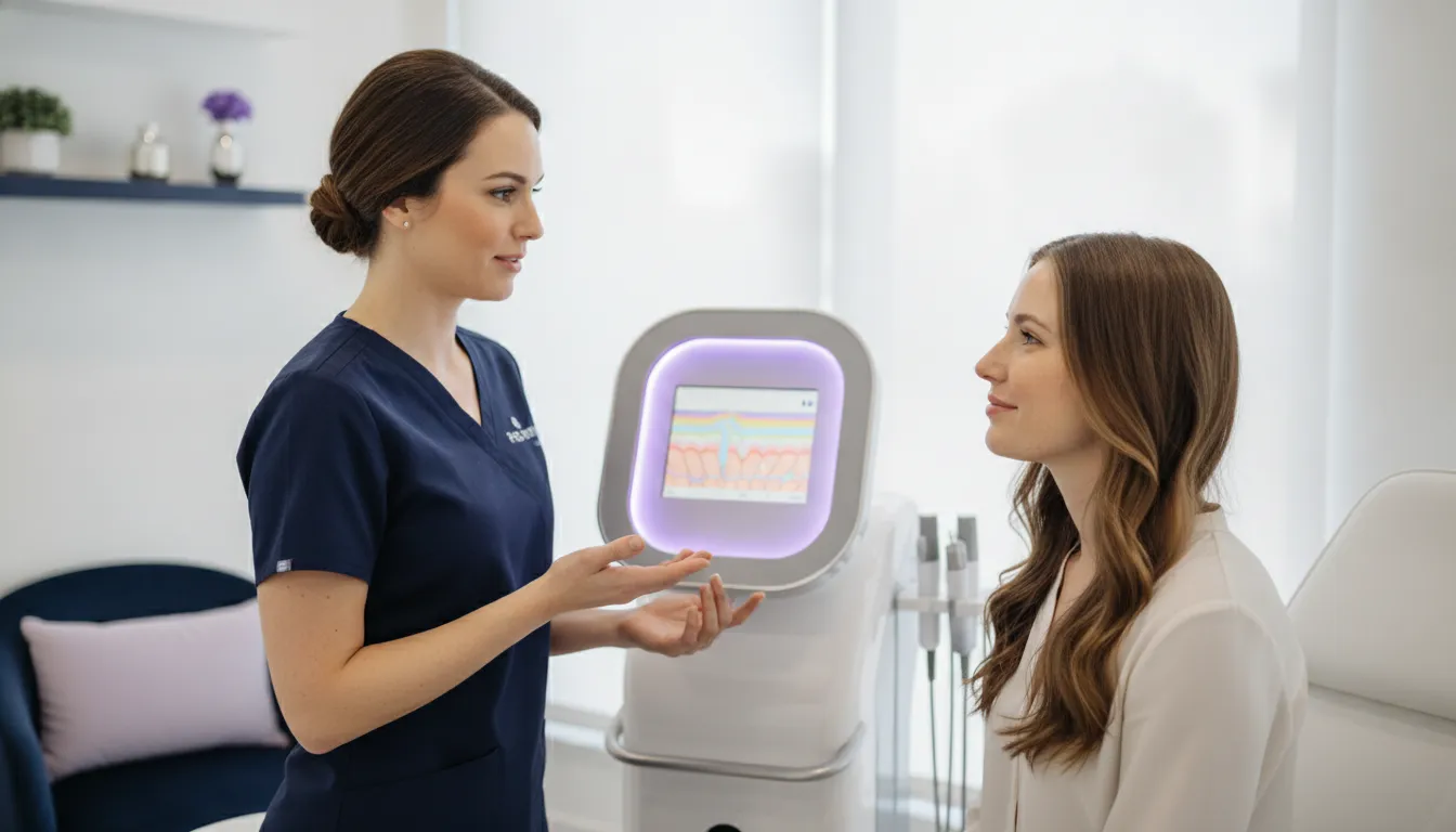
A skin image is a record of what a camera captured under particular lighting and positioning. By itself, it does not explain what those visible details mean or how they relate to a client’s goals.
In an aesthetic practice, AI skin analysis uses software to assess features in captured skin images and organize them as observations for professional discussion. It can add a structured reference to imaging, but it is not a diagnosis and does not replace clinical judgment.
Request a product tour to learn about available imaging workflows.
For example, Emage describes ImagePro skin imaging systems as using analysis data to inform care-plan recommendations aligned with a practice’s offerings.
The distinction matters: image capture creates the visual record; software interpretation adds another way to review it. Understanding what each contributes, and where its limits lie, is the starting point for using imaging responsibly in consultation.
What Is AI Skin Analysis and How Does It Differ from Traditional Skin Imaging?
AI skin analysis is software-assisted review of captured skin images. It organizes visible features into observations that a practitioner can use as another reference during a consultation. The camera and lighting create the images; analysis software then processes those images. Neither step, by itself, establishes a medical diagnosis or replaces professional judgment.
Traditional skin imaging gives a practitioner a visual record to inspect, discuss, and potentially compare over time. Depending on the system, images may be captured under different lighting conditions to make certain surface features easier to observe. AI adds software-based organization or measurements to that image-based workflow. Emage describes its ImagePro systems as using AI skin-analysis data to support a care plan aligned with a practice’s offerings, while available capabilities vary by model. Practices can review the ImagePro skin imaging systems to see how the product line is presented.
| Approach | What it contributes | Limitations |
|---|---|---|
| Traditional visual review | A professional examines captured images and discusses visible features with the client. | Interpretation depends on image conditions and the practitioner’s review; a photograph is only one source of context. |
| Skin imaging | Creates a visual record, sometimes with different lighting modes. For example, Emage lists normal, parallel-polarized, cross-polarized, and UV imaging modes for SUNLITE. | Images can show appearance, not the full history, symptoms, or other context needed for professional assessment. |
| AI-assisted image analysis | Software processes captured images to present structured observations that can support discussion and planning. | Outputs depend on the image and system; they should be interpreted in context, not treated as a diagnosis or guaranteed finding. |
These approaches can complement one another rather than compete. AI does not make the original capture more informative if the image is unclear, nor can software supply a client’s history or goals. A review of dermatology AI research notes that performance can be affected by image quality and by missing information such as medical history and risk factors. Those caveats matter when deciding how much weight to give any software-generated observation.
For practices, the practical distinction is what happens after capture: a conventional image remains a reference for human review, while AI-assisted analysis may organize visual information for a more structured conversation. Both require a practitioner to explain what is visible, ask relevant questions, and decide what belongs in the client’s plan. For background on the broader imaging workflow, see this broader guide to professional skin analysis.
Which Skin Features Can AI Imaging Help Practitioners Assess?
Depending on the system and its analysis features, image-based tools may help practitioners review visible details such as pores, wrinkles, pigmentation, redness, and skin texture. These are visual observations for consultation and documentation, not evidence of a disease and not a diagnosis. The features a system can assess, and the way it presents them, vary by model.
Research illustrates why those distinctions matter. One study evaluated automated grading of facial signs including forehead and periorbital wrinkles, nasolabial folds, and diffuse redness. In that study, automated cheek-pore grading correlated moderately with dermatologist assessments, while pigmentation grading correlated weakly, particularly for the darkest skin tones. These results describe one system and study population; they should not be treated as proof of another product’s performance. Read the study’s methods and findings.
For an aesthetic practice, an image can provide a consistent visual reference to discuss a client’s concerns and goals. An analysis label or score should be interpreted alongside the actual image and the professional’s assessment. Lighting, image quality, capture conditions, and the specific model’s capabilities all shape what can be observed. If a feature is unclear or does not fit the client’s experience, do not treat the software output as definitive.
Skin-tone representation deserves particular care. A published evaluation found weak correlation for pigmentation grading, especially for the darkest skin tones, in its study. That is a reason to ask vendors what populations and conditions were included in validation and to avoid assuming that a result transfers equally across skin tones or devices. This study is not a performance finding about any specific Emage system.
Models can also differ in the imaging modes and workflows they offer. For example, Emage describes ImagePro HYBRID imaging features separately from other systems in its lineup. Review the specifications for the particular model under consideration rather than assuming every platform captures or analyzes the same features. Whatever the setup, use the images to support a careful professional conversation, not to promise a clinical conclusion or outcome.
How Can AI Skin Analysis Support Patient Consultations?
A captured image can give the practitioner and client a shared visual reference during a consultation. Rather than relying only on a mirror or memory, the practitioner can point to features the system makes visible, ask what the client notices, and hear which concerns matter most to them. This can make the discussion more specific, while keeping the client’s goals and the practitioner’s assessment at the center.
Use the analysis as a prompt for conversation, not a verdict. Ask whether a displayed observation matches the client’s concerns, explain what the image can and cannot show, and consider it alongside the client’s history and an appropriate professional assessment. Research on dermatology AI has identified limitations in model performance across skin tones and uncommon conditions, which is a reason to discuss outputs cautiously rather than treat them as definitive. The findings concern studied AI models, not a validation of any particular aesthetic imaging product (study of the Diverse Dermatology Images dataset).
When moving from observations to options, make the reasoning understandable. Emage describes ImagePro systems as using AI skin-analysis data to support a care plan aligned with a practice’s offerings. The practitioner should explain why an option may be relevant, invite questions, and document what the client agrees to consider. A model-specific example is ImagePro HYBRID; available features and workflows vary across systems. Any printed or digital analysis can support education, but it does not replace professional interpretation or establish a diagnosis.
For a returning client, a prior capture can also serve as a reference point for discussing changes over time. Emage describes facial recognition on SUNLITE as supporting returning-client progress charting and comparison. Before interpreting differences, check that the images are suitable to compare and discuss what else may have changed since the earlier visit. A comparison documents what is visible in the captured images; it does not by itself show why a change occurred or establish that a service caused it.
Set expectations before capturing or sharing images. Explain how the practice will use them, who may access them, how they will be stored, and whether the client has a choice about participation. Follow the practice’s consent and privacy procedures, and avoid promising a particular result. The technology is most useful in consultation when it helps both parties ask clearer questions and make a considered, documented plan, with the practitioner responsible for the interpretation.
Integrating AI Skin Analysis into Patient Intake and Treatment Planning
A consistent process makes image-based observations easier to interpret alongside the details a patient shares. Treat the system as a documentation and consultation aid: its output can inform a professional discussion, but it is not a diagnosis or a substitute for clinical judgment.
- Start with intake, history, and goals. Ask what the patient would like to address and record relevant background through the practice’s usual intake process. Keep those details available during review rather than treating an image as a complete account of a person’s skin or concerns. A review of dermatology AI research notes that image quality and missing clinical history can limit interpretation, reinforcing the need to consider context with any image-based output (scoping review of dermatology AI).
- Explain the process and obtain consent. Before capturing images, explain what the imaging session is for, how images and results will be used, and who may review them. Follow the practice’s consent, privacy, and retention procedures. Give the patient an opportunity to ask questions and proceed only when they understand and agree.
- Use a repeatable capture routine. Follow the system’s instructions for positioning, lighting, and framing, and keep the setup consistent when images will be compared over time. Recheck the capture if the image is blurred, poorly framed, or otherwise not comparable. Do not treat a change in image conditions as a change in the patient’s skin. Imaging capabilities vary by model; for example, ImagePro SUNLITE is described as supporting 3D imaging and follow-up procedures.
- Review observations with professional context. Have the appropriate practitioner review the images and any software-generated observations alongside the intake information and the patient’s stated goals. Describe outputs as visual references, not definitive findings. If an image or concern calls for assessment beyond the system’s non-diagnostic role, use the practice’s established professional process rather than relying on an AI result.
- Discuss options and document the agreed plan. Explain which observations informed the conversation, answer questions, and discuss suitable options within the practitioner’s scope and the practice’s offerings. Record the patient’s priorities, the options discussed, and the plan they agree to, including any follow-up. Emage describes ImagePro AI skin-analysis data as supporting care-plan recommendations aligned with practice offerings; the practitioner remains responsible for the discussion and plan.
- Compare follow-up images carefully. At a later visit, repeat the agreed capture routine and review images side by side with the prior record. Note differences as observations, accounting for capture conditions and other context; avoid promising a particular result. For more on the foundations of a professional imaging workflow, see this broader guide to professional skin analysis.
What Should Practices Look for When Evaluating AI Skin Imaging Systems?
Start by defining the job the system needs to do. An imaging platform can support consultation, visual documentation, and discussion of care options, but it should not be treated as a diagnostic device or a substitute for professional judgment. Emage describes its ImagePro systems as using AI skin-analysis data to support care-plan recommendations aligned with a practice’s offerings. That describes a consultation aid, not proof of a diagnosis or guaranteed result. Compare the workflow you need with the functions listed for the specific model, rather than assuming every system has the same capabilities.
- Clarify intended use. Ask the vendor what the system is designed to capture, analyze, and report, and what it is not intended to do. Confirm that staff understand the boundary between visual observations and clinical assessment.
- Review capture conditions. Ask which lighting and imaging modes are available on the model under consideration, how staff select them, and what conditions must stay consistent between visits. Different illumination can reveal different visual information, but useful comparisons still depend on repeatable capture.
- Test repeatability in your workflow. Have staff follow the same positioning and capture steps with different operators. Ask how the software handles image quality and what it does when a capture is unsuitable for comparison. A polished report is not a substitute for a consistent process.
- Inspect model-specific outputs. Request a demonstration of the actual model’s features, analysis views, and patient-facing materials. Emage’s ImagePro skin imaging systems include multiple configurations; verify what is included rather than extrapolating from another model or a general product-line description.
- Ask for validation evidence and its limits. Request information about the data and population used to evaluate the relevant outputs, including representation across skin tones, and ask how performance was tested in real practice conditions. In one published study of automated grading of facial signs, correlation with dermatologist assessments varied by sign; pigmentation grading was weakly correlated, particularly for the darkest skin tones. The study illustrates why a single accuracy claim may not answer the practical question. Review the study methods and findings, and ask vendors for evidence specific to their system.
- Confirm operations and data handling. Ask what patient data and images are collected, where they are stored, who can access them, how long they are retained, and what consent and deletion controls are available. Review the vendor’s written terms with your practice’s privacy lead. Also assess staff training, report usability, support, and compatibility with your existing intake and documentation process.
Use a live demonstration to test these questions against your own patient journey and staffing needs. To review the available models and workflows, request a product tour.
Frequently Asked Questions
How accurate is AI skin analysis?
There is no single accuracy score that applies to every system, image, or skin feature. Image quality, capture conditions, the feature being assessed, and the software’s validation all matter. A study comparing automated facial-sign grading with dermatologists found that agreement varied by sign, so treat image outputs as reference points for professional discussion, not definitive measurements or a substitute for clinical judgment. Read the study.
Does skin imaging provide a medical diagnosis?
No. Emage Medical describes ImagePro systems as imaging tools for consultation and documentation, not as diagnostic devices. Their images or analysis can help organize observations and support a conversation, but they do not establish a medical diagnosis. If a patient has a skin concern that needs clinical assessment, refer it to an appropriately qualified clinician rather than relying on an imaging result.
How can a practice check whether a system works across skin tones?
Ask the vendor what populations and skin tones were represented in validation, which features were tested, and whether results were assessed in real-world conditions. Do not assume that performance on one feature applies to another. Research has identified limitations in dermatology AI performance on darker skin tones and emphasized the need for diverse validation. See the study on diverse dermatology images.
How can we compare repeat images more consistently?
Use a documented capture routine. Keep the device and lighting mode, camera distance, face position, and framing as consistent as practical, and note differences that could affect the image. Compare like with like and review the images alongside the patient’s history and goals. A change in lighting, pose, or image quality can affect what appears visible, so avoid treating a visual difference alone as proof of clinical change.
Ready to Review ImagePro Options?
A product tour can give your team a practical way to compare ImagePro models and consider how an imaging workflow may fit your aesthetic practice. To discuss the options, request a product tour when convenient.