

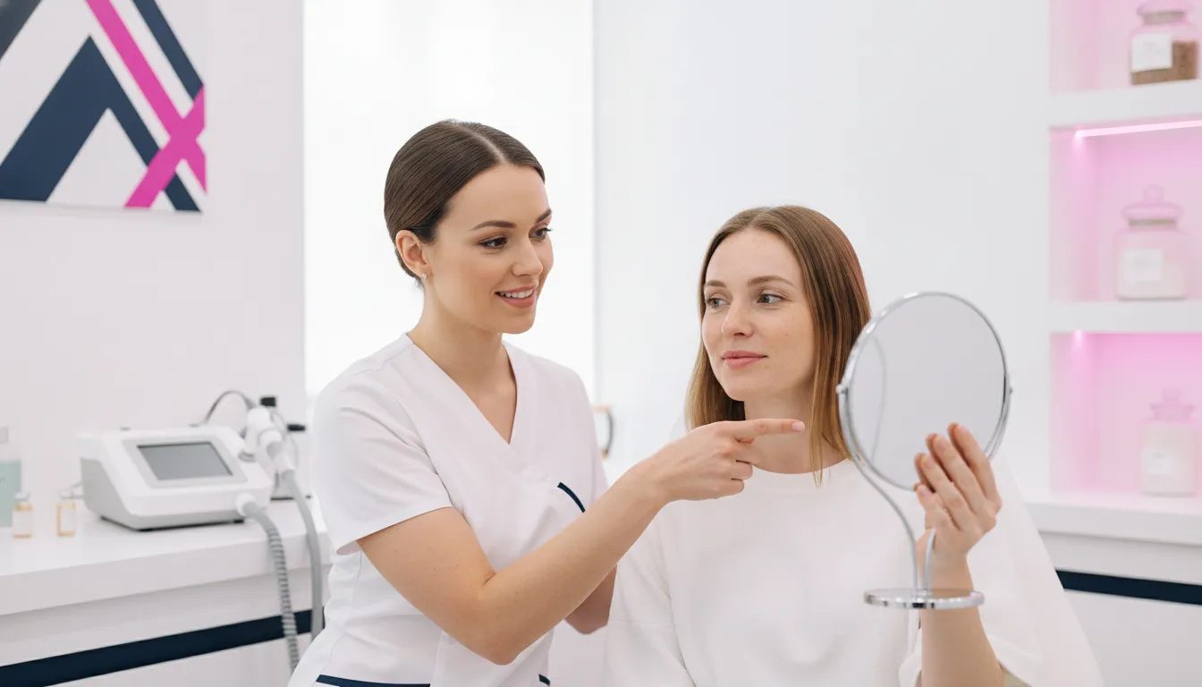
Microneedling before and after photos can make a consultation more concrete. But they should be treated as part of a documented patient story, not as a promise of identical results. Patient selection, protocol, provider technique, skin condition, photography conditions, and follow-up timing all affect what a viewer sees. For an aesthetic practice, the value is in creating a repeatable way to discuss appearance changes while preserving clinical judgment and realistic expectations.
Request a product tour to review professional microneedling equipment for your practice.
For aesthetic practices, microneedling before and after documentation can illustrate changes in texture, fine lines, scars, or overall skin appearance. Photographs are most useful when captured consistently and reviewed with treatment records and clinical evaluation.
That distinction helps providers explain what an image can show and what it cannot prove. A consistent workflow, informed consent, and privacy-conscious records support more credible consultations. The right professional microneedling pens and devices can complement that process, but the first step is understanding how to document and interpret results responsibly.
What Can Microneedling Before and After Results Actually Show?
A useful comparison should show a relevant change under comparable conditions, not simply a different photograph. Microneedling uses controlled needle penetration to create micro-injuries. The skin’s repair response may influence appearance over time. The pace and visible effect depend on the patient and treatment plan. Provider technique, skin condition, device settings, and follow-up timing also matter.
Depending on the patient’s concern and the provider’s assessment, photographs may help document smoother-looking texture, less prominent pores, or a gradual softening in the appearance of fine lines. Practices may also discuss the appearance of atrophic acne scars, uneven pigmentation, and stretch marks. These categories should not be treated as interchangeable indications, and a photograph should not be presented as proof of a guaranteed outcome.
Interpret each concern on its own terms
Texture and pore appearance can be affected by lighting, hydration, makeup, camera distance, and skin preparation. Fine lines may look different as remodeling progresses, but age, sun exposure, skin quality, and maintenance habits also influence what is visible. Acne scar documentation requires attention to scar type and depth. A comparison image cannot establish that every structural component of a scar has improved.
Hyperpigmentation needs especially careful interpretation. Exposure, inflammation, skincare, and other procedures may alter skin tone independently of microneedling. Stretch marks may be an appropriate consultation topic, but outcomes are variable. The practical goal is to define a patient-specific objective and document the context around it rather than promise uniform fading.
Use evidence and variability to frame the conversation
Clinical literature describes microneedling applications and ongoing questions about protocols, but evidence and outcomes are not identical across every concern or patient. A responsible review pairs standardized photographs with treatment records, clinical evaluation, and informed consent. It also distinguishes an early post-procedure appearance from a later remodeling result. Practices evaluating a professional microneedling pen system should keep those documentation limits in mind when assessing technology and setting patient expectations.
How Many Sessions Do Patients Typically Need to See Visible Changes?
Session planning should provide a realistic framework rather than a promised deadline. A practice can explain when early changes may be assessed, when remodeling may become more apparent, and when the treatment plan should be reviewed. The schedule remains individualized because the concern, skin response, protocol, device settings, treatment area, and follow-up timing influence what appears in photographs and in person.
- Start with the concern and treatment objective. Skin rejuvenation, texture concerns, acne scarring, photoaging, and stretch marks may not follow the same response pattern. The clinical assessment should determine whether microneedling is appropriate and what a reasonable course could look like.
- Use a recovery interval as a planning reference. Many professional protocols space sessions several weeks apart, allowing the skin to recover and giving the practice time to evaluate the response. This is a reference point, not an instruction for every patient. The provider may adjust timing based on the concern, intensity and settings used, healing, and clinical history.
- Explain what a multi-session course means. A course of three sessions spaced several weeks apart can take multiple months. That example helps patients understand why one immediate photograph may not represent a later appearance. Some concerns may be discussed as requiring additional sessions, but the number should remain a provider-led planning decision rather than a guarantee.
- Schedule assessment after the final session. A useful review point may be several weeks after the final treatment, when the practice can compare standardized photographs and clinical observations. Immediate redness, swelling, or other temporary changes can make an early image misleading.
- Review maintenance only when appropriate. Follow-up should consider the patient’s response, skin condition, care factors, and ongoing goals. The provider should decide whether further treatment is clinically appropriate and document the reasoning.
Clear documentation turns a timeline into a useful consultation tool. It helps the care team explain why visible changes may develop gradually and why results can differ between patients, even when the same technology is used.
Request a product tour to compare microneedling platforms and discuss your practice workflow.
How Should Practices Set Expectations During Consultations?
A productive consultation should make the limits of before-and-after photography clear. A photograph can document a change in appearance under defined conditions. But it cannot establish that microneedling alone caused the change or predict the same response for another patient. Treatment discussions should remain specific to the patient’s concern, goals, medical history, proposed protocol, and follow-up plan.
Practices should describe downtime accurately without presenting it as universal. Temporary erythema, sensitivity, swelling, or other reactions can vary with the treatment area, device settings, skin characteristics, and patient response. RF microneedling may involve a different recovery profile because it combines needling with radiofrequency energy. A qualified treating professional should handle discussions of potential complications, contraindications, and follow-up.
Discuss the treatment horizon, not a guaranteed endpoint
Patients may need more than one session, and visible changes can develop gradually as the skin remodels. Maintenance may also be part of a longer-term plan. Longevity depends on the treated concern, patient characteristics, protocol, follow-up timing, and ongoing care. Practices should use before-and-after examples to explain variability, not to imply that another patient will have the same result.
- What specific concern is being evaluated, and what would count as a meaningful but realistic change?
- How many sessions might be considered, what interval is clinically appropriate, and when will progress be reviewed?
- What downtime and possible adverse effects should the patient understand before consenting?
- How will treatment settings, photographs, clinical notes, and follow-up timing be documented?
- Will maintenance be considered, and what factors could affect the durability or visibility of change?
Consistent records help a practice interpret images responsibly. Consultation photos should be considered alongside clinical assessment, consent, treatment details, and follow-up observations rather than used as standalone proof of efficacy.
How Can You Document Before and After Results More Reliably?
Well-documented photographs can make a consultation more concrete, but they are one part of an outcome record. Differences in lighting, angle, expression, skin preparation, camera settings, healing stage, and follow-up timing can make results appear greater or smaller than they are. Use a repeatable process that gives the clinical team and patient a fair basis for comparison.
- Define the documentation purpose before treatment. Record the patient’s concerns, treatment area, relevant history, planned protocol, and the outcome being evaluated. Patient education, clinical follow-up, internal quality review, and marketing may require different consent and privacy controls.
- Standardize the photo environment. Use consistent lighting, camera position, distance, background, focus, and orientation. Keep the patient’s expression, hair placement, and facial position as consistent as reasonably possible. Avoid filters, aggressive retouching, or changes that could obscure the condition being evaluated.
- Set intervals that reflect the treatment plan. Photograph the baseline before treatment, document relevant follow-up visits, and avoid treating an immediately post-procedure image as the final result. The exact timing should follow the provider’s clinical evaluation and the patient’s response.
- Pair images with treatment records. Note the device or modality, treatment settings used under the provider’s protocol, dates, treatment areas, patient-reported experience, observed skin changes, and relevant aftercare or adverse events.
- Obtain informed consent and protect privacy. Explain how images will be used, who may see them, how they will be stored, and whether permission may be withdrawn where applicable. Keep identifying information separate from images when possible and restrict access to the consented purpose.
A clinical study protocol available through ClinicalTrials.gov illustrates why standardized photography, fixed positioning, consistent conditions, and recorded treatment details matter when comparing baseline and follow-up images. A practice may use a similar operational mindset without treating a study protocol as a universal clinical instruction.
Used together, standardized photography, informed consent, treatment records, and clinical evaluation create a more credible account of progress than a carefully selected image alone. A professional skin imaging system may also help organize visual discussions, but imaging remains a consultation and documentation aid, not a substitute for clinical judgment.
What Is the Difference Between Standard and RF Microneedling?
Both modalities use controlled needle penetration to support a wound-healing response and dermal remodeling. Standard microneedling relies on the mechanical action of sterile needles creating controlled micro-injuries. RF microneedling adds radiofrequency energy delivered through the needles, creating a second treatment component that combines mechanical needling with thermal energy.
The added energy does not guarantee better outcomes. Patient selection, treatment parameters, provider training, informed consent, and follow-up remain central to appropriate care. A practice should compare platforms according to the services it plans to offer, the qualifications of its team, its protocol requirements, and the documentation workflow it can support.
These are planning distinctions, not promises. Practices should establish protocols within their clinicians’ training and applicable requirements, document informed consent, and set expectations around variability rather than comparing devices through a single before-and-after image.
For practices evaluating a professional RF platform, Emage Medical describes Cyto-RF RF microneedling technology as FDA-cleared. FDA clearance applies to the specific device and its cleared uses; it does not guarantee a particular patient’s result. Product discussions should connect the selected modality to the patient’s condition, goals, clinical history, and an appropriately documented plan.
How Does Professional Equipment Support a Consistent Patient Workflow?
Equipment should fit into the full patient journey, not sit apart from consultation, treatment planning, documentation, and follow-up. A consistent workflow gives licensed providers a clearer way to prepare patients, record what was performed, and discuss what changes may be observed over time. It does not guarantee a particular result. Patient selection, provider qualifications, protocol, skin condition, photography conditions, and follow-up timing all influence how images should be interpreted.
Connect consultation, treatment, and records
Standardized intake and treatment records can help a practice document the clinical context behind each image. Records may include the patient’s stated concerns, relevant observations, the treatment plan, and the follow-up interval. Consistent photography, informed consent, privacy protection, and appropriate clinical evaluation should accompany those records.
Practices evaluating the professional microneedling pen and device category can consider how a platform fits existing room setup, documentation habits, staff responsibilities, and the services they are qualified to provide. CytoPen NG and CytoPen S2 are presented by Emage Medical as professional microneedling options. Product claims should be reviewed against the specific product page and its intended use.
Build repeatability through training and implementation
Repeatable protocols are easier to implement when staff understand the equipment, the practice’s approved procedures, and the boundaries of their roles. Product demonstrations, virtual training, installation, and clinical education can help a practice introduce technology with a defined implementation plan. The goal is not to make every patient identical. It is to reduce avoidable variation in preparation, communication, recordkeeping, and follow-up.
The CytoPen NG professional microneedling pen page can help qualified buyers review one platform in the context of a broader device portfolio. A product tour can then focus the discussion on device fit, intended workflow, training questions, and the information a practice needs before making a purchasing decision.
Request a product tour to discuss a professional microneedling workflow with Emage Medical.
Frequently Asked Questions
When should a practice take after photographs?
Capture follow-up images at a consistent interval after treatment, rather than immediately when temporary swelling or redness may affect appearance. Use the provider’s clinical assessment to select the timing, and keep lighting, camera position, expression, background, and patient preparation as consistent as possible. Review photographs alongside treatment records and clinical evaluation.
How long does it take to see results from microneedling?
Visible changes may develop gradually, and the timing varies by patient, concern, protocol, device settings, and follow-up interval. A practice should explain when it plans to review progress without promising a specific outcome or deadline. An early image should not automatically be treated as the final result.
How many microneedling sessions might a patient need?
The number of sessions is individualized. It depends on the concern being evaluated, the patient’s skin response, the selected protocol, treatment settings, and the provider’s clinical judgment. Practices can discuss a possible course during consultation, then reassess the plan using standardized photographs, treatment notes, and follow-up observations.
What makes a microneedling before and after photo more credible?
Credible documentation uses consistent photography conditions, informed consent, clear records, and an appropriate follow-up interval. It also explains what else may have influenced the appearance, including skincare, other procedures, lighting, healing, and patient factors. A photo can support a conversation, but it cannot prove efficacy or guarantee another patient’s result.
Sources: NCBI Bookshelf overview of microneedling; ClinicalTrials.gov study protocol record.